
3D images of cancer cells in the body: medical physicists from Halle present a new method
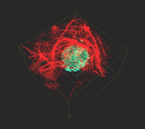
Photo: Jan Laufer
Bringing tumour cells to light: medical physicists at the Martin-Luther-University Halle-Wittenberg (MLU) have developed a new method with which detailed three dimensional images of inside the body can be produced. This may help, for example, to examine the development of cancer cells in the body more closely. The group of researchers is presenting its development in the “Communication Physics” specialist journal from the Nature Publishing Group.
Doctors and researchers need an even better understanding of cancer cells and their properties to be able to target the treatment of cancer. In research, individual cancer cells are often analysed in test tubes first before the findings can then be checked in living organisms. “We want to spy on the cancer cells directly inside the body though and find out how they spread, how they work and how they react to innovative therapies,” says medical physicist Prof. Dr. Jan Laufer from MLU. He has specialised in the field of so-called photoacoustic imaging, a method with which high-resolution, three dimensional images of inside the body can be produced with the help of ultrasonic waves generated by laser beams.
“The problem is that tumour cells are transparent. This makes it difficult to examine tumours in the body using an optical method,” explains Laufer. With his working group he has developed a new method to solve this problem: this involves the scientists implanting a specific gene into the cancer cells’ genome first of all. “The phytochrome protein, which originally comes from plants and bacteria, is produced by the gene in the cells. It acts as a light sensor there,” continues Laufer. In the next step, the researchers fire short light pulses onto the tissue in the body. They use a laser with two different wavelengths for this. The light pulses are absorbed inside the body and converted into ultrasonic waves. These waves can then be measured outside the organism and two images of inside the body can be produced on the basis of these data. “What is special about the phytochrome proteins is that they change their structure and therefore also their absorption properties depending on the laser beams’ wavelength. As a result, the amplitude of the optically induced sound waves in the tumour cells changes. All the other tissue components, for example the blood vessels, don’t have this property, their signal remains constant,” explains Laufer. By calculating the difference between the two images, a high-resolution three dimensional image is produced of the tumour cells that doesn’t have a disturbing background contrast.
The development by the medical physicists from Halle can be applied to a whole range of photoacoustic examinations: besides cancer research, the method is also suitable for observing the way cellular and genetic processes work inside living organisms.
About the study:
J. Märk et. al, Dual-wavelength 3D photoacoustic imaging of mammalian cells using a photoswitchable phytochrome reporter protein. Communication Physics 2018, 1, 3. DOI: 10.1038/s42005-017-0003-2
Contact partner:
Prof. Dr. Jan Laufer
Medical Physics
Phone: +49 345 55-25400
E-Mail: jan.laufer.ignore@physik.uni-halle.de
Web: http://www.physik.uni-halle.de/fachgruppen/medphysik/